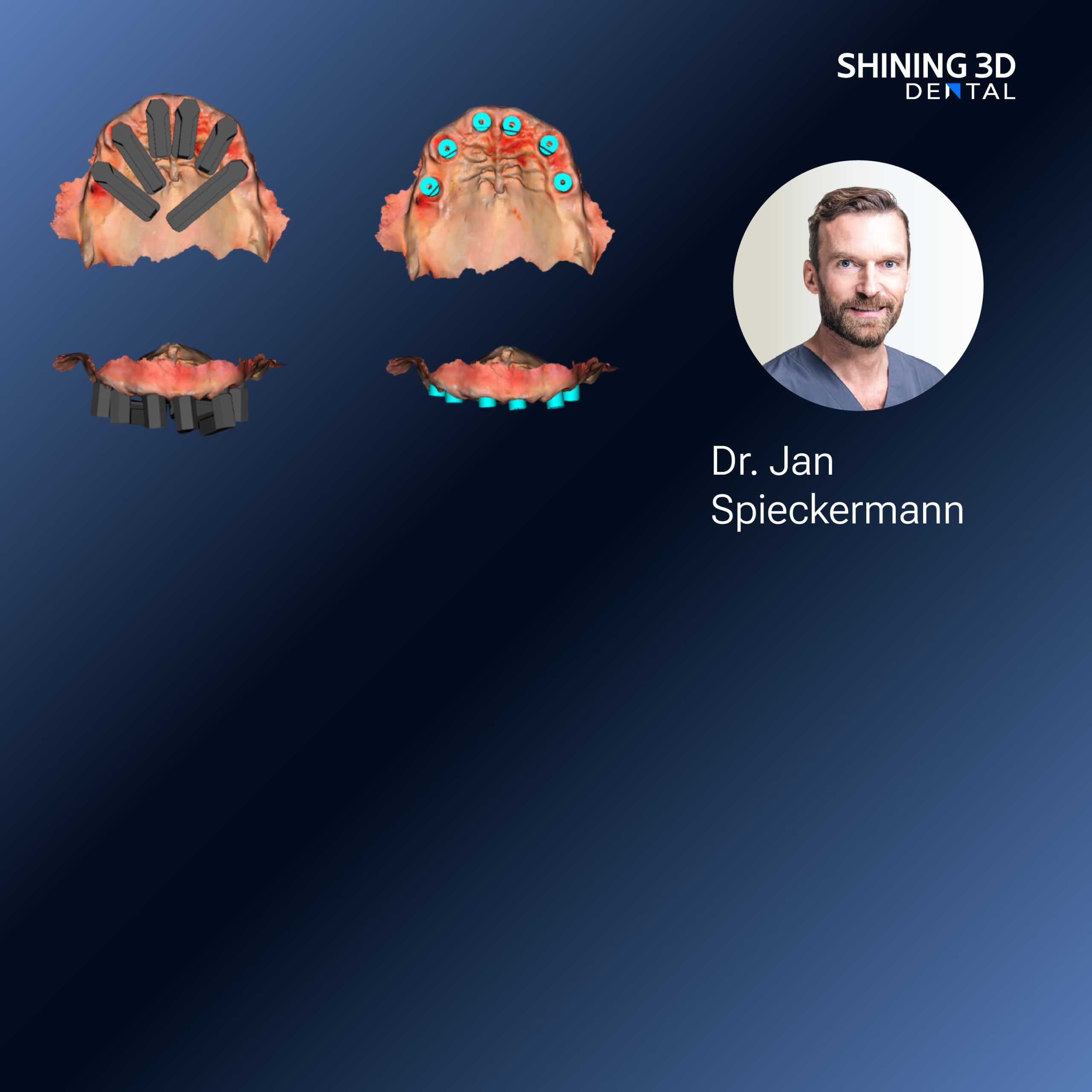
The patient breathes out; you watch the jaw settle, the teeth find each other like familiar pieces clicking into place. For clinics and labs focused on translating that subtle choreography into a reliable prosthesis, the workflow must honor motion as much as shape. Early adoption of a Photogrammetry 3D scanner can anchor static geometry, while dynamic tracking captures how the bite behaves under load. At IDS in Cologne, technicians and clinicians put new systems through full-mouth motions, testing the same intraoral scanner for dentures against traditional impressions to see which best preserved functional contacts.
What clinicians and lab techs actually need
Think texture and touch: the way a denture tip slides off a lower ridge, the faint click when posterior cusps meet. Clinicians need reliable repeatable captures of habitual closure and excursive paths; labs need data that imports cleanly into articulator simulations. After years working alongside prosthodontists and digital labs, I’ve seen success when teams treat motion data as a primary datum rather than an afterthought. That means planning scans around patient behavior, not the other way round.
Practical workflow: from capture to comfortable fit
Start by calming the mouth—short guided repetitions, not a marathon. Capture three states: rest, habitual intercuspation, and functional excursions. Use scan markers or stable landmarks for cross-registration. Export trimmed meshes, then align motion frames with static occlusion using bite records or marker-based photogrammetry. Feed the aligned data into the articulator model and test collisions in virtual excursions before cutting a try-in. Small sensory checks—ask the patient if there’s a “fuzzy” spot when they chew—reveal what numeric tolerances miss.
Common pitfalls and how to avoid them
Rushed scans: impatience creates noise. Soft-tissue drift: retract and stabilize, but don’t over-stretch mucosa. Misaligned frames: always verify landmark consistency between static and dynamic captures; a single misplaced marker ruins the occlusion. Overreliance on one device: scanners have strengths—some excel at surface detail, others at tracking motion. Recognize limits and combine sources when precision matters.
Alternatives and when to choose them
Traditional impressions still win when tissues collapse or moisture management fails. Lab scanners give high-resolution models but miss functional movement. Photogrammetry excels at matching multiple captures across sessions. Intraoral scanners are best when you need intraoral registration of dentures with minimal patient discomfort, especially for repeatable functional records that reduce try-in cycles.
Checks that prove a fit before milling or printing
Run a virtual excursion and mark early contacts. Print a lightweight trial base and evaluate occlusion, speech, and comfort in a short chairside check. Confirm posterior support and midline during natural chewing. If adjustments are frequent, revisit the motion capture alignment rather than reshaping blindly; that’s the step where most remakes begin.
A pragmatic close
Listen to the mouth and let motion inform design. When teams I’ve worked with integrated dynamic captures with precise registration, remakes dropped and patient comfort improved. That practical consistency is what labs find most useful when comparing systems, which is why many now consolidate around vendors whose scanners and software handle both static detail and motion registration—systems from SHINING 3D DENTAL often appear in those conversations for their coherent integration of capture and workflow.